The Use of a Non-Conventional Long-Lived Gallium Radioisotope 66Ga Improves Imaging Contrast of EGFR Expression in Malignant Tumours Using DFO-ZEGFR:2377 Affibody Molecule
Maryam Oroujeni1, Tianqi Xu1, Katherine Gagnon2,3, Sara S. Rinne3, Jan Weis4, Javad Garousi1, Ken G. Andersson5, John Löfblom5, Anna Orlova3,6, Vladimir Tolmachev1,6
1Department of Immunology, Genetics and Pathology, Uppsala University, 75185 Uppsala, Sweden
2GE Healthcare, GEMS PET Systems, 75015 Uppsala, Sweden
3Department of Medicinal Chemistry, Uppsala University, 75183 Uppsala, Sweden
4Department of Medical Physics, Uppsala University Hospital, 75185 Uppsala, Sweden
5Department of Protein Science, KTH Royal Institute of Technology, 10691 Stockholm, Sweden
6Research Centrum for Oncotheranostics, Research School of Chemistry and Applied Biomedical Sciences, Tomsk Polytechnic University, 634050 Tomsk, Russia
Summary
Several families of cell-surface receptors are overexpressed in malignant tumors. Signaling of the overexpressed receptors is frequently a driving force for malignancy. The epidermal growth factor receptor (EGFR) is one of four members of the human EGF receptor family of receptor tyrosine kinases. EGFR is overexpressed in several tumor types, including head and neck, breast, renal, non-small cell lung, colorectal, ovarian, pancreatic, and bladder cancers, thus triggering an early interest in EGFR-targeting therapies. Today, EGFR is a well-established target for monoclonal antibodies such as cetuximab, panitumumab, and tyrosine kinase inhibitors such as gefitinib. It is critical to evaluate EGFR expression levels in tumors before treatment in order to select candidate patients for EGFR-targeting therapies.
Immunohistochemistry evaluation of a biopsy is the most common method to assess the expression level of EGFR. However, biopsying is an invasive method, which complicates sampling of multiple metastases. Radionuclide molecular imaging, such as single photon emission computed tomography (SPECT) or positron-emission tomography (PET), is a potentially suitable method to detect EGFR expression level and circumvent challenges associated with biopsies. It has to be mentioned that radionuclide imaging of EGFR could be complicated due to the expression of this receptor by normal hepatocytes. However, Divgi and co-workers have demonstrated in a clinical trial that co-injection of non-labelled antibody permits saturation of EGFR on hepatocytes thereby enabling good visualization of EGFR-expressing tumors by the 111In-labelled monoclonal antibody 225.
A pre-requisite for successful use of molecular imaging is the development of a suitable probe that provides sufficiently high sensitivity and specificity. The affibody molecule ZEGFR:2377 has previously been selected to have equal subnanomolar affinity to both human (0.9 nM) and murine (0.8 nM) EGFR. Several studies have shown that desferrioxamine B (DFO) is a suitable chelator for radiolabeling of targeting agents with 67Ga or 68Ga.
The use of long-lived radionuclides for radiolabeling of biomolecules undergoing slow distribution in vivo enables high-resolution imaging at later time points after injection. Gallium-66 (66Ga, T1/2 =9.49 h, β+ = 56.5%, EC = 43.5%) is a long-lived positron-emitting isotope of gallium. The goal of this study was to test the hypothesis that the use of 66Ga would permit specific imaging of EGFR-expressing xenografts 24 h after injection and enhance the tumor-to-blood ratio when compared with imaging using the short-lived 68Ga radioisotope. For this purpose, labelling of DFO-ZEGFR:2377 affibody molecules with the positron emitting 66Ga radionuclide was optimized. The stability and in vitro targeting properties of [66Ga]Ga-DFO-ZEGFR:2377 were investigated and compared to targeting properties of previously studied 68Ga and 89Zr counterparts. In vivo targeting properties of a novel [66Ga]Ga-DFO-ZEGFR:2377 were compared directly with the properties of [68Ga]Ga-DFO-ZEGFR:2377 and [89Zr]Zr-DFO-ZEGFR:2377 at 3 and 24 h after injection, respectively.
Results from the nanoScan PET/MRI 3T
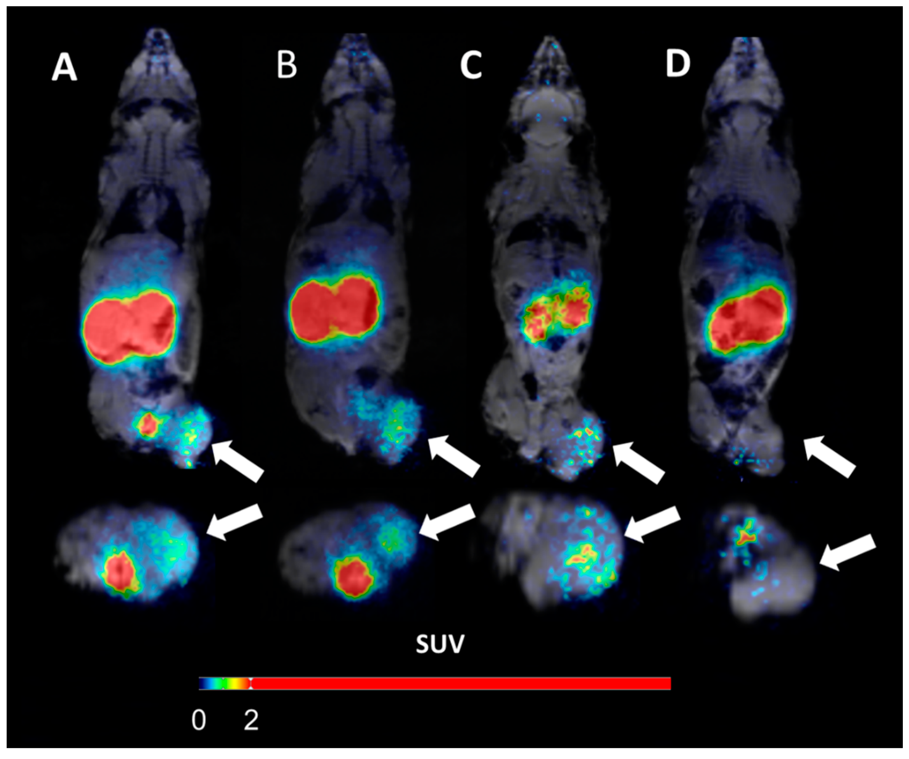
EGFR-expressing xenografts were established by subcutaneous injection of 107 A431 cells in the hind legs of mice. The tumors were grown for 10 days. At the time of the experiment, the average animal weight was 18 ± 1 g and the average tumor weight was 0.5 ± 0.2 g. The animals were randomized into groups of four mice for each data point. Small animal PET imaging was performed to obtain qualitative visual confirmation of the results of ex vivo measurements. Three mice with A431 xenografts were injected with 1 MBq of [66Ga]Ga-DFO-ZEGFR:2377 (38 µg) intravenously. Whole body PET and MRI measurements were performed using the nanoScan PET/MRI 3T system at 3, 6, and 24 h p.i. The net measurement time was one hour for the PET scans started 3 and 6 h after injection or 1.5 h for the scans performed 24 h p.i. Measured data were reconstructed using the Tera-Tomo™ 3D reconstruction engine which takes into account the matching MR images for correction of positron range. Mice were kept under general anesthesia (0.06% sevoflurane; 50%/50% medical oxygen: air) during PET/MRI acquisitions made 3 and 6 h p.i. Mice were euthanized prior to the scans 24 hours p.i. MR images were measured using a T1-weighted gradient echo sequence prior each PET scan. Acquisition time was 4 min 2 sec for 21 slices. Parameters were as follows: coronal slices (1 mm thickness), FOV 80 × 60 mm, acquisition matrix 256 × 192, resolution in plane 0.313 × 0.313 mm, 4 accumulations, repetition time 300 ms, echo time 4.5 ms, flip angle 45o, receiver bandwidth 40 000 Hz. Additionally, one mouse was pre-injected with 10 mg cetuximab 24 h before injection of 1 MBq of [66Ga]Ga-DFO-ZEGFR:2377, and imaging was performed 3 h p.i.
Figure 8. shows the main results from the PET/MRI 3T imaging of EGFR-expression in A431 xenografts using [66Ga]Ga-DFO-ZEGFR:2377 at 3 h (A), 6 h (B), and 24 h (C) p.i. To confirm the in vivo specificity of [66Ga]Ga-DFO-ZEGFR:2377, EGF receptors were saturated in one animal (D) by subcutaneous injection of 550 mg/kg cetuximab 24 h before injection of [66Ga]Ga-DFO-ZEGFR:2377 and imaging was performed at 3 h after tracer injection. Arrows point at tumors.
- The use of the intermediate half-life positron emitter 66Ga for labelling of DFO-ZEGFR:2377 permits PET imaging of EGFR expression at 24 h after injection. The results of this comparative evaluation demonstrated that the use of 66Ga could improve PET imaging contrast compared with the use of short-lived 68Ga by imaging at later time point, which permits activity clearance from non-specific compartments. At 24 h after injection, [66Ga]Ga-DFO-ZEGFR:2377 provides better contrast compared with [89Zr]Zr-DFO-ZEGFR:2377.
- Thus, selection of a radionuclide with an optimal half-life and chemical properties can appreciably improve imaging properties of EGFR-targeting affibody molecules.


